21 Jul Vein Occlusions
Vein occlusions are commonly seen in a retinal specialist’s office and are second only to diabetic
retinopathy in frequency of problems affecting the blood vessels of the eye.
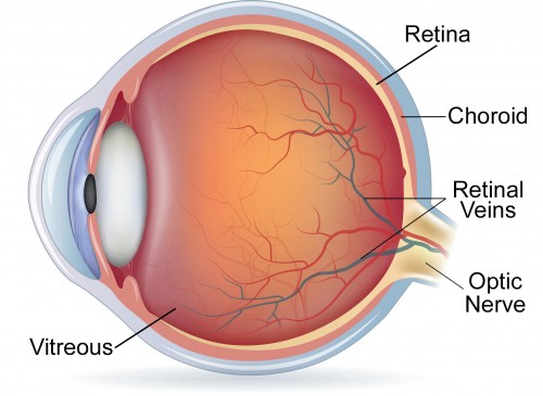
There are two types of vein occlusions: the branch retinal vein occlusion and the central retinal vein occlusion. The blood vessels of the retina are like a tree. There is a major trunk, the central retinal artery, which supplies branches which get smaller and smaller. Then the blood returns through branches of the retinal vein which get bigger and bigger until it finally terminates in the central retinal vein.
Both the central retinal artery and vein enter the back of the eye through the optic nerve. Branch retinal vein occlusion occurs when there is a blockage of one of the branches of the retinal vein.
Central retinal vein occlusions occur where the main trunk is still in the optic nerve in the wall of they just before it exits the eye itself.
As you can imagine central retinal vein occasions result in poorer vision than branch retinal vein occlusions just like a blockage in your home’s main sewage line results in more water backup and damage than a branch line of your sewage line.
Poor vision results from either a disruption of the blood supply as the blood has a difficult time draining from the retina, or from leakage of fluid from damaged blood vessels.
For a long time the only treatment that could be offered the patient was laser. Laser is effective when there is a rare complication where the blood supply is so disrupted that new vessels grow and can bleed. This is a rare event. The more common need for laser was to treat the leakage of fluid from damaged blood vessels. Unfortunately, laser although helpful was not dramatically useful in improving vision.
All this changed in the last 10 years with the development of drugs, which reverse the leakage in the damage blood vessels. These drugs are injected into the eye itself and are the same drugs which are used to treat age related macular degeneration and diabetic retinopathy.
When you visit a retinal specialist, a study which examines blood flow is performed (fluorescein angiography) and a study which maps out swelling of the retina (ocular coherence tomography.) Both of these studies help the retina specialist determine if treatment is necessary.