What is a Retinal Detachment
“Retina” is the name for the nerve cells that line the inside of the back of the eye. The retina acts like a film in a camera receiving the light that is focused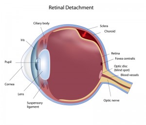 by the structures in the front of the eye and forming an image that is transmitted to the brain where vision is completed. To function normally, the retina must remain attached to the inside of the back of the eye. Situated in front of the retina is the vitreous body, a large gel-like structure that is adherent to the retina throughout the first four or five decades of life.
What is a PVD?
With aging, the gel vitreous liquefies and eventually it collapses. When the vitreous body collapses it pulls away from the retina. This is known as posterior vitreous detachment, or PVD. This event happens to almost everyone if they live long enough. In most people vitreous separates away from the retina cleanly without any problems. In some people, however, there are problems, collectively known as anomalous PVD.
Sebag J: Anomalous PVD – a unifying concept in vitreo-retinal diseases. Graefes Arch Clin Exp Ophthalmol 242:690-698, 2004.
Sebag J: Vitreous Anatomy, Aging, and Anomalous Posterior Vitreous Detachment. In: Encyclopedia of the Eye (Dartt, Besharse, Dana, eds.) Elsevier, Oxford, Vol. 4, pp. 307-315, 2010]
Diagnosis of Retinal Detachment
One of the potential manifestations of anomalous PVD is traction upon a site where vitreous is so firmly adherent to the retina that it breaks the retina. This usually occurs in an area of the retina that is not important to vision. However a broken retina can lead to blindness if it becomes a retinal detachment. This occurs when fluid accumulates under the retina and lifts the retina off the inside of the back of the eye. Nearsighted eyes, head or eye trauma, and eyes that have undergone cataract surgery are at greater risk of retinal detachment A retinal detachment is identified by an examination with special viewing instrument called an indirect ophthalmoscope and/or using ultrasonography.
Symptoms of Retinal Detachment
The symptoms of a retinal detachment are floaters, flashing lights, and a dark curtain.
Floaters are hair-like, fly-like, grey, linear structures that move with eye movement and keep moving a little once the eye stops moving. PVD is the cause of floaters. If the PVD pulls on the retina it can cause a retinal tear. Flashing lights can appear with either a PVD or a retinal tear. These lights are usually bright, white, linear, and located off to the side of the visual field. If fluid accumulates under the retina then vision is lost in the area corresponding to the area of detached retina. This is usually up above, down below, or off to the side in the field of vision. There is usually a curved line that demarcates the area of vision loss from the area of normal vision. It is important to have this symptom evaluated and treated as soon as possible. Treatment can be an office procedure called pneumatic retinopexy, removal of the vitreous body that caused the retinal detachment, called vitrectomy, or a sewing a plastic-like band to the outside of the eye, called scleral buckle.
J. Sebag, MD, FACS, FRCOphth, FARVO
Retina Specialist
Huntington Beach, CA 92647